The bone defect reconstruction process can use hydroxyapatite is osteoconductive and can retain the original biocompatible shape to enhance hydroxyapatite with osteogenic proteins. To analyze the most appropriate concentration of hydroxyapatite nanoparticles using the MTT Assay method and to test the viability of osteoblast cells after being given hydroxyapatite nanoparticle (nHA) derived from unam snail shells. The fabrication of hydroxyapatite nanoparticles from unam snail shells using a mechanical-chemical combination method. Osteoblast cells are obtained from Calvaria rats after being cultured in DMEM. Viability tests of osteoblast cells were done using the MTT Assay method and repeated three times, and then results were measured using an Elisa reader. Viability of osteoblast cells in nHA 1,25 mg/ml (164,60 % ± 0,096), nHA 1,5 mg/ml (151,72 % ± 0,176), nHA 1,75 mg/ml (90,55 % ± 0,243), nHA 2 mg/ml (74,23 % ± 0,301) respectively. ANOVA test shows p < 0,05. IC50 value of hydroxyapatite nanoparticle from the unam snail’s shells to viability osteoblast cells is 2,23 mg/ml. Less concentration of hydroxyapatite nanoparticles tends to increase the viability of osteoblast cells. 1,75 mg/ml and below hydroxyapatite nanoparticles derived from unam snail shells are not toxic to osteoblast cells.
This is an Open Access article, distributed under the terms of the Creative Commons Attribution 4.0 International License (http://creativecommons.org/licenses/by/4.0/), which permits unrestricted use, distribution and reproduction in any medium or format, provided the original work is properly cited.
Bone Graft, Viability Test, Hydroxyapatite, Osteoblast Cells, Unam Snail Shells
1. Introduction
The alveolar bone surrounding the tooth socket is renovated continuously. The adjusted balance between bone resorption and bone formation is maintained by different types of cells and signaling mechanisms. In susceptible individuals, the inflammatory response to bacteria can initiate the destructive process of periodontitis, leading to loss of connective tissue and bone and apical migration of epithelial junctions. While disruption of subgingival microbial biofilms and periodontal resolution of inflammation can be achieved with non-surgical therapy, regeneration usually cannot be expected, thus leading to a reduction in periodontium and possible residual alveolar bone damage. In more advanced cases of periodontitis, the tooth loses support and is chipped or extracted. Therefore, the clinical need for alveolar bone regeneration appears to improve the long-term prognosis of teeth with periodontitis. In addition to bone loss due to chronic inflammation, bone regeneration may be necessary to correct defects caused by other things, including trauma, tumor resection, or congenital and developmental abnormalities
[1]
Larsson L, Decker A. M, Giannobile W. V. Regenerative Medicine for Periodontal and Peri-Implant Disease. J of Dent Research. November 2015; 95(3): 1-12.
[1].
Mechanical therapy, antimicrobial therapy, or surgical therapy may be used to treat periodontal disease. Mechanical therapy such as scaling, root planing, and polishing
[2]
Jin L, Lamster I, Greenspan J, Pitts N, Scully C, Warnakulasuriya S. “Global burden of oral diseases: emerging concepts, management, and interplay with systemic health,” Oral Diseases 2016; 22(7): 609–619.
[3]
Ryan ME. Nonsurgical approaches for the treatment of periodontal diseases. Dent Clinics North Am. 2005; 49: 611-36.
[2, 3]
cannot restore the attachment of periodontal tissue to the tooth, but only prevents the development of periodontal disease and reduces symptoms. Guided Tissue Regeneration (GTR) and bone grafts (bone grafts) aim to restore the structure and function of the periodontal tissue in patients with periodontitis
[4]
Deporter DA. Periodontal disease part II: Overview of treatment modalities. Can Fam Physician. 1988; 34: 1391-2.
[4]
which includes the repair of cementum, bone, and periodontal fibers in the hope of preventing adhesions. between teeth and soft tissue, alveolar bone regeneration, and reduction of soft tissue inflammation
[5]
Liang Y, Luan X, Liu X. Recent advances in periodontal regeneration: A biomaterial perspective. Bioactive Materials. 2020; 5(2): 297-308.
[6]
Mohammad T, Zeenat I, Javed A, Sanjula B, Sushama T, Zulfiqar A, et al. Treatment modalities and evaluation models for periodontitis. International J of Pharm Investigation. July 2012; 2(3): 106-122.
[5, 6]
.
An ideal bone graft material should be non-toxic, non-irritating, mechanically resistant, able to adapt to the body (biocompatible), capable of being absorbed by biological tissues (bioresorbable), osteoinduction, osteoconduction, able to resemble real bone, simple to use, and economical
[7]
Elly M, Mohamad R, Asti M. Used Bone Graft for Periodontal Defect Treatment. JKGUI. 2003; 10: 520-526.
[7]
.According to the source, bone graft materials used for periodontal regeneration can be divided into natural transplants (autograft, allograft, xenograft) and synthetic materials (alloplastic) that are widely used in reconstructive periodontal surgery to fill periodontal defects so that periodontal tissue regeneration can occur
[8]
Grado de GF, Keller L, Idoux-Gillet Y, et al. Bone Substitutes: A Review of Their Characteristics, Clinical Use, and Perspectives for Large Bone Defects Management. J Tissue Engineering. 2018; 9: 1-18.
[9]
Bayani M, Torabi S, Shahnaz A, Pourali M. Main properties of nanocrystalline hydroxyapatite as a bone graft material in the treatment of periodontal defects. A review of the literature. Biotechnology & Biotechnological Equipment. 2017; 31(2): 215-20.
[8, 9]
these materials have benefits and drawbacks when used to treat existing bone defects. With these limitations, research in the field of biomaterials can be stimulated by modifying the production of synthetic biomaterials, whose properties resemble those of natural bone and can be precisely measured and controlled. Among the various synthetic bone graft materials currently available, hydroxyapatite is a synthetic material (alloplastic) that has demonstrated great potential as a bone defect-filling material due to its biocompatibility and chemical similarity to the hydroxyapatite found in bone tissue
[9]
Bayani M, Torabi S, Shahnaz A, Pourali M. Main properties of nanocrystalline hydroxyapatite as a bone graft material in the treatment of periodontal defects. A review of the literature. Biotechnology & Biotechnological Equipment. 2017; 31(2): 215-20.
[10]
Astuti A, Maria U. Sintesis dan karakterisasi komposit hidroksiapatit dari tulang ikan lamuru (sardilnella longiceps)-kitosan sebagai bone filler. JF FIK UINAM. 2017; 5(1): 9-15.
[9, 10]
Hydroxyapatite is found in nature such as in limestone and in bio-inorganic materials such as fish scales, teeth, snail shells, clams, coral, or egg shells. Snail shells from the mollusk phylum, especially gastropods, can be used for the synthesis of hydroxyapatite because they contain a lot of calcium in their shells. One example is the Unam snail (Volegalea cochlidium). Carpenter and Niem stated that the golden snail has a hard and dense shell with a relatively large shell size (up to 15 cm in length) and contains 95-99.9% calcium carbonate interspersed with 0.1-5% organic protein. The harder the shell, the higher the calcium carbonate (CaCO3) content. So, this snail shell is suitable for use as a source of calcium hydroxyapatite
[11]
Morris JP, Backeljau T, Chapelle G. Shells from Aquaculture: A Valuable Biomaterial, Not a Nuisance Waste Product. Reviews in Aquaculture. 2019; 11: 42-57.
[12]
Hou Y, Shavandi A, Carne A, et al. Marine Shells: Potential opportunities for extraction of functional and health-promoting materials. Critical Reviews in Environmental Science and Technology. 2016; 46(11-12): 1047-1116.
[11, 12].
In general, hydroxyapatite is synthesized from snail shells via the precipitation method, which involves the chemical reaction between calcium and phosphorus ions under a pH- and temperature-controlled solution
[13]
White M, Cheljava M, Fried B, et al. The Concentration of Calcium Carbonate in Shells of Freshwater Snails. Amer Malac Bull. 2007; 22: 139-42.
[13]
This method has several flaws, including many variables that can influence the results of hydroxyapatite synthesis, such as the initial concentration of reactants, the pH of the reaction, and the synthesis temperature, and it yields a product consisting of large crystals. Recently, the sol-gel method was introduced, which produces nanoparticle-sized hydroxyapatite powder with homogeneous grains, a high degree of crystallinity, and a low processing temperature
[14]
Agrawal K, Singh G, Puri D, Prakash S. Synthesis and characterization of hydroxyapatite powder by sol-gel method for biomedical application. J of Minerals & Materials Characterization & Engineering. 2011; 10(8): 727-34.
[14]
Utilizing existing shell waste, the researcher is interested in conducting research on the shells of the Unam snail (Volegalea cochlidium) based on the background information provided. To achieve optimal and safe results in the treatment of bone defects caused by periodontal diseases using bone graft material derived from the unam snail shell, a biocompatibility test must be performed on the bone graft material. The degree of cytotoxicity, mutagenicity, and cancer-causing potential of a material determines its biocompatibility when placed in the body
[15]
Ganachari SV, Bevinakatti AA, Yaradoddi JS, Anapurmath NR, Hunashyal AM, Shettar AS. Rapid synthesis, characterization, and studies of hydroxyapatite nanoparticles. Adv Mater Sci Res. 2016; 1(1): 9-13.
[16]
Sirait M, Sinulingga K, Siregar N, et al. Characterization of hydroxyapatite by cytotoxicity test and bending test. J. Phys.: Conf. Ser. 2022; 2193: 1-8.
[15, 16]
The purpose of this study was to determine the level of toxicity of a substance so that it can be accepted by the host, is not carcinogenic, and does not cause allergic reactions. This is the initial phase of testing a material that will be a candidate for bone graft material in the field of Periodontics in dentistry
[16]
Sirait M, Sinulingga K, Siregar N, et al. Characterization of hydroxyapatite by cytotoxicity test and bending test. J. Phys.: Conf. Ser. 2022; 2193: 1-8.
[16].
2. Material and Methods
This research is a laboratory experimental study on osteoblast cell culture with a post-only control group design. The population of this study was osteoblast cells in 24 well plates, each of which was 1x10⁶ and the sample for this research was the hydroxyapatite nanoparticles of the Unam Snail shell (Volegalea cochlidium).
The research sample was divided into six treatment groups for 24 hours. Calculation of the number of samples in each group used in this study is to use Federer's (1963) simple laboratory experiment formula, namely:
(n-1) (r-1) ≥ 15
(4-1) (r-1) ≥ 15
3 (r-1) ≥ 15
r ≥ 3
where:
n = total of treatment groups
r = number of samples per group
From the calculation results, the sample size obtained for each treatment group is 3 times the replication.
Free variables were the concentration of the Unam snail shell hydroxyapatite nanoparticles and the size < 100nm. The dependent variable was osteoblast cell culture viability. Controlled variables were laboratory procedures, the contact time of the test material with cell culture 24 hours, and the number of cells in the culture.
It required 1 kilogram of unam snail shell that has been cleaned of its meat, while the remaining portions can be cleaned with a pry tool. The snail shells were then washed with distilled water until they were clean, crushed with a hammer, and ground with a mortar and pestle. The ball mill was activated by pressing the main switch (main switch on). The hood of the grinding chamber was opened button and then the hood down was pressed. The open command until the automatic lock appeared.
The sol-gel method is a wet chemical synthesis method that does not require a high pH value and refers to the low-temperature chemical precursor method. In the sol-gel method, a solution of ammonium is heated to 60°C, and then 181 grams of EDTA are added and stirred until dissolved. To obtain pure hydroxyapatite with a Ca/P ratio of 1.67, the powder was mixed with an appropriate amount of diammonium hydrogen phosphate and stirred at 90oC until the Ca/P ratio reached 1.67.
This study performed the MTT assay on the 24-hour group, including a control group without cell media, control cells without treatment, and cells treated at 1 mg/ml; 1.5 mg/ml; 1.75 mg/ml, and 2 mg/ml. A 570-595 nm wavelength Elisa reader is used to read the results.
Data Analysis
Data from each examination were analyzed statistically with a significance level (α = 0.05), starting with the data normal ty test using the Shapiro-Wilk method followed by a one-way analysis of variance test (ANOVA), to determine differences in cytotoxicity between all treatment groups.
3. Results
In the Integrated Laboratory of the University of North Sumatra, hydroxyapatite powder was produced by synthesizing the shells of Unam snails (Volegalea cochlidium) using a ball mill with sol-gel, to obtain homogeneous and nanometer-sized hydroxyapatite particles. In addition, the osteoblast cell viability test was conducted after exposure to hydroxyapatite from the unam snail shell at the UPT Integrated Laboratory and CDAST Technology Innovation Center, University of Jember, which was divided into a control group and concentration groups of 1.25 mg/ml; 1.5 mg/ml; 1.75 mg/ml; 2 mg/ml using the MTT assay method, then optical density (OD) is obtained and readings are conducted with an Elisa reader. The mechanical production of hydroxyapatite nanoparticles from snail shells (Volegalea cochlidium) using a ball mill with stainless steel balls at 500 rpm for one hour, two hours, three hours, and 1500 rpm for one hour, two hours, and three hours. The average particle size of the unam snail shell is 40 nm. After the ball milling process, sol-gel is followed by a chemical method.
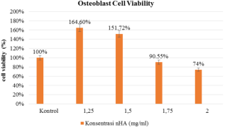
In Table 1 the group (K4) with a nHA concentration of 1.25 mg/ml had the highest average percentage of osteoblast cell viability, which was 164%. This demonstrates that a concentration of 1.25 mg/ml of nHA can stimulate the growth of osteoblast cells without being toxic.
Table 1. Optical Density (OD) of the treatment group after administration of hydroxyapatite nanoparticles from the unam snail shell in osteoblast cell culture.
Group (K)
nHA concentration
Viability Percentage Mean ± SD
K1
2 mg/ml
74,23 % ± 0,301
K2
1,75 mg/ml
90,55 % ± 0,243
K3
1,5 mg/ml
151,72 % ± 0,176
K4
1,25 mg/ml
164,60 % ± 0,096
The absorbance value (Optical Density) in the MTT assay within 24 hours was lowest in the group (K1) with a concentration of 2 mg/ml = 74.23% ± 0.301 and highest in the group (K4) with a concentration of 1.25 mg/ml = 164.60% ± 0.096, according to the results of research on osteoblast cells treated with hydroxyapatite nanoparticles of unam snail shells.
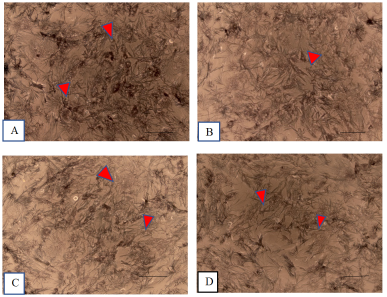
Microscopic view of osteoblast cells after treatment with hydroxyapatite nanoparticles of Unam snail shells through an inverted microscope at concentrations of 1.25 mg/ml; 1.5 mg/ml; 1.75 mg/ml; 2 mg/ml reveals very dense formazan fiber bonds; this indicates the number of osteoblast cells that are alive after treatment in the MTT assay after 24 hours. Increasing test material concentration decreases the frequency of formazan fiber bonds (Figure 1)
Figure 1. Microscopic view of osteoblast cells after being treated with nHA (A) 1.25 mg/ml; (B) 1.5 mg/ml; (C) 1.75 mg/ml; and (D) 2 mg/ml (Red arrow = formazan fiber, black line = 0.040 µm. Magnification 20 x).
Optical Density (OD) data for each concentration within 24 hours can be seen using the formula: Cell viability (%) = (OD treatment/OD control) x 100% to determine the percentage of osteoblast cell viability after administration of hydroxyapatite nanoparticles from snail shells unam (Volegalea cochlidium). The results were obtained with the highest viability in the concentration group (K4) and the lowest viability in the concentration group (K1) within 24 hours (Figure 2).
Figure 2. Graph of osteoblast cell viability after administration of hydroxyapatite nanoparticles from unam snail shells (Volegalea cochlidium).
The resulting data was then subjected to a normality test with Shapiro-Wilk because the number of samples was less than 50. The normality test results for all data yielded a p-value <0.05, which means the resulting data was not normally distributed. The data were tested for homogeneity to see the homogeneity of the data, the test results showed p = 0.002 (p <0.05) which indicated that the data obtained was not homogeneous. Then a non-parametric test was performed with Kruskall Wallis to assess the median difference between concentrations, the test results obtained p = 0.014 (p <0.05) indicating a significant difference in the absorbance value at each concentration. The Mann-Whitney test is used to determine the difference in the median of the 2 groups if the dependent variable's data scale is ordinal or interval/ratio but not normally distributed. The results obtained in each group were significantly different.
In Table 2 the Kruskal Wallis test yielded a significant value of 0.014 (<0.05), indicating that there is a difference between the absorbance values of each group of unam snail shell hydroxyapatite concentrations. To determine the difference in the number of viable cells between concentration groups, the Mann-Whitney test was conducted. The average absorbance value between concentration groups varied significantly (< 0.05), indicating that the higher the concentration of nHA administered, the fewer viable osteoblast cells were detected.
Table 2. Kruskall Wallis and Mann-Whitney analysis of osteoblast cell viability.
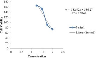
IC50 was calculated using a regression analysis between the percentage of viability and the concentration of nHA. Based on the graph, the equation for the regression line is y = (132.92)x + 336.27 with an IC50 value of 2.23 mg/ml, which indicates that the maximum concentration of nHA can stimulate osteoblast cell proliferation by 50%. The value of the determinant of the correlation coefficient (R2) is close to 1 (0.9267 or 92%), indicating a close relationship between the concentration variable and viability.
Figure 3. IC50 calculation regression line equation.
4. Discussion
This study utilized hydroxyapatite from unam snail shell waste (Volegalea cochlidium) obtained from the Serdang Bedagai Sea Area of North Sumatra and then processed at the UPT Integrated Laboratory of the University of North Sumatra with a ball mill to produce nanoparticles. The PSA (Particle Size Analyzer) test yielded a particle size of 0.040 µm or 40 nm at 500 rpm in 1 hour. This site is considered a nanoparticle because the average particle size is < 100 nm.
A chemical process was conducted at 900°C using the sol-gel method to obtain homogeneous hydroxyapatite. The obtained results were in the form of powdered hydroxyapatite with a composition of 39% Ca and 18.5% P. This is theoretically compatible where the calcium and phosphorus content (in % by weight) in the formation of hydroxyapatite is respectively 39.6 % and 18.45 % with a Ca/P ratio of 1.6753
[17]
Yoyada, Novendi. Viability Test Of Bovine Tooth Graft On cell Culture Fibroblast BHK 21 With MTT Assay Method. Thesis FKG Unair. 2015: 5-24.
[17]
The viability test of osteoblast cells after administration of hydroxyapatite nanoparticles in concentrations of 1.25 mg/ml, 1.5 mg/ml, 1.75 mg/ml, and 2 mg/ml.
To achieve healing through periodontal regeneration, bone graft materials derived from autograft, allograft, xenograft, and alloplastic are used to treat periodontal disease accompanied by bone damage. One of the requirements for these materials is that they are biocompatible, which means that they have no toxic or detrimental effects on the biological environment, both locally and systemically. To determine a material's toxicity level, a clinical trial known as the viability test is conducted. The viability test is used to evaluate the biological effects of a substance and determine the cell's response to external stimuli by measuring the ability of a cell to survive after a specific exposure. This test is typically performed to determine a material's biocompatibility
[18]
Anuar A, Salimi MNA, Zulkal M, Daud M, Yee YF. Characterizations of hydroxyapatite (HAp) nanoparticles produced by sol-gel method. Intl Conf Adv Mater Eng Technol. 2013: 3587-3590.
[19]
Bandyopadhyay SG. Bone as a Collagen-hydroxyapatite Composite and its Repair, Trends Biomater, Artif. Organs 2008; 22(2): 112.
[20]
Aminatun, Muhammad HI, Jan A, et al. Synthesis and Characterization of Nano-Hidroxyapatite/ Chitosan/ Carboxymethyl Cellulose Composite Scaffold. J Int Dent Med Res 2019; 12(1): 31-37.
[18-20]
.
This study employed an enzymatic test with MTT assay reagent (2-(4,5-dimethyl-2thiazolyl)-3, 5-diphenyl-2H tetrazolium bromide) to determine the viability of a sample.
[21]
Shokrzadeh M, Modanloo M. An overview of the most common methods for assessing cell viability. J Res Med Dent Sci 2017; 5(2): 33-41.
[21]
By examining the metabolic activity of a cell through coulometry, this test is utilized more frequently by researchers because it is sensitive, faster, and more accurate in determining a cell's viability. Additionally, this test is used to determine the cytotoxicity of toxic substances and other medical agents.
Cell culture is the process by which a cell is grown under controlled conditions and is derived from cells that have been separated from their original tissue, namely cell lines or cell strains. Because animal cells grow more slowly than pathogens such as fungi and bacteria, the culture conditions for this cell culture must always be aseptic. In this study, osteoblast cells from mouse calvaria were utilized. Quantitative information on osteoblast cell culture is obtained by calculating the percentage of viable cells in the culture. In each group, Optical Density (OD) measurements yielded these outcomes
[19]
Bandyopadhyay SG. Bone as a Collagen-hydroxyapatite Composite and its Repair, Trends Biomater, Artif. Organs 2008; 22(2): 112.
[19]
In this study, osteoblast cell culture was used to evaluate the viability of hydroxyapatite nanoparticles derived from unam snail shells due to the role of osteoblast cells in the formation of the bone matrix during bone remodeling and osteogenesis to promote tissue regeneration
[13]
White M, Cheljava M, Fried B, et al. The Concentration of Calcium Carbonate in Shells of Freshwater Snails. Amer Malac Bull. 2007; 22: 139-42.
[14]
Agrawal K, Singh G, Puri D, Prakash S. Synthesis and characterization of hydroxyapatite powder by sol-gel method for biomedical application. J of Minerals & Materials Characterization & Engineering. 2011; 10(8): 727-34.
[13, 14].
This study was conducted in vitro with osteoblast cells isolated from the calvaria of mice. Each concentration group (K1) 2 mg/ml (74.23%), (K2) 1.75 mg/ml (90.55%), (K3) 1.5 mg/ml (151.72%), and (K4) 1.25 mg/ml (164.60%) averaged viable osteoblast cells.
The IC50 value is the inhibitory concentration of 50% of the test substance, hydroxyapatite nanoparticles of unam snail shells, on osteoblast cells, as determined by a regression equation between the percentage of viability and the concentration of hydroxyapatite nanoparticles. With a coefficient value of 0.9267 mg/ml, or close to 1, the maximum concentration of nHA can stimulate osteoblast cell proliferation by 50%, resulting in a value of approximately 1. The IC50 value of hydroxyapatite nanoparticles from the shell of the unam snail (Volegalea cochlidium) is 2.23 mg/ml, as determined by the study. Based on these results, it can be concluded that the percentage of viable cells increases as the concentration of the test material decreases.
The MTT Assay method was used to determine the viability of osteoblast cells in this study within 24 hours; at the lowest concentration of 1.25 mg/ml, a cell viability percentage of 164.60% was obtained, indicating that many osteoblast cells are viable, but not yet dividing. The number of cells that undergo proliferation and differentiation is known. Therefore, it is necessary to conduct additional in-vitro studies with a longer observation period to determine the number of cells that undergo proliferation and differentiation, as well as the type of cells formed. This is important information, as the nHA extracted from snail shells in this study will be considered for use as a bone graft material in the field of Periodontics. Long-term, it is also necessary to conduct additional research on experimental in-vivo animals to determine the compatibility of the unam nanoparticles derived from snail shells
[22]
Chang M, Wu J, Liao H, Chen Y, Kuo C. Comparative Assessment of Therapeutic Safety of Norcantharidin, N-farnesyloxy-norcantharimide, and N-farnesyl-norcantharimide against Jurkat T Cells Relative to Human Lymphoblast. Med 2016; 95:
[22]
.
5. Conclusion
The effective concentration of hydroxyapatite nanoparticles is less than 2 mg/ml. Less concentration of hydroxyapatite nanoparticles tends to increase the viability of osteoblast cells. 1,75 mg/ml and below hydroxyapatite nanoparticle derived from unam snail’s shells is not toxic to osteoblast cells.
Larsson L, Decker A. M, Giannobile W. V. Regenerative Medicine for Periodontal and Peri-Implant Disease. J of Dent Research. November 2015; 95(3): 1-12.
[2]
Jin L, Lamster I, Greenspan J, Pitts N, Scully C, Warnakulasuriya S. “Global burden of oral diseases: emerging concepts, management, and interplay with systemic health,” Oral Diseases 2016; 22(7): 609–619.
[3]
Ryan ME. Nonsurgical approaches for the treatment of periodontal diseases. Dent Clinics North Am. 2005; 49: 611-36.
[4]
Deporter DA. Periodontal disease part II: Overview of treatment modalities. Can Fam Physician. 1988; 34: 1391-2.
[5]
Liang Y, Luan X, Liu X. Recent advances in periodontal regeneration: A biomaterial perspective. Bioactive Materials. 2020; 5(2): 297-308.
[6]
Mohammad T, Zeenat I, Javed A, Sanjula B, Sushama T, Zulfiqar A, et al. Treatment modalities and evaluation models for periodontitis. International J of Pharm Investigation. July 2012; 2(3): 106-122.
[7]
Elly M, Mohamad R, Asti M. Used Bone Graft for Periodontal Defect Treatment. JKGUI. 2003; 10: 520-526.
[8]
Grado de GF, Keller L, Idoux-Gillet Y, et al. Bone Substitutes: A Review of Their Characteristics, Clinical Use, and Perspectives for Large Bone Defects Management. J Tissue Engineering. 2018; 9: 1-18.
[9]
Bayani M, Torabi S, Shahnaz A, Pourali M. Main properties of nanocrystalline hydroxyapatite as a bone graft material in the treatment of periodontal defects. A review of the literature. Biotechnology & Biotechnological Equipment. 2017; 31(2): 215-20.
[10]
Astuti A, Maria U. Sintesis dan karakterisasi komposit hidroksiapatit dari tulang ikan lamuru (sardilnella longiceps)-kitosan sebagai bone filler. JF FIK UINAM. 2017; 5(1): 9-15.
[11]
Morris JP, Backeljau T, Chapelle G. Shells from Aquaculture: A Valuable Biomaterial, Not a Nuisance Waste Product. Reviews in Aquaculture. 2019; 11: 42-57.
[12]
Hou Y, Shavandi A, Carne A, et al. Marine Shells: Potential opportunities for extraction of functional and health-promoting materials. Critical Reviews in Environmental Science and Technology. 2016; 46(11-12): 1047-1116.
[13]
White M, Cheljava M, Fried B, et al. The Concentration of Calcium Carbonate in Shells of Freshwater Snails. Amer Malac Bull. 2007; 22: 139-42.
[14]
Agrawal K, Singh G, Puri D, Prakash S. Synthesis and characterization of hydroxyapatite powder by sol-gel method for biomedical application. J of Minerals & Materials Characterization & Engineering. 2011; 10(8): 727-34.
[15]
Ganachari SV, Bevinakatti AA, Yaradoddi JS, Anapurmath NR, Hunashyal AM, Shettar AS. Rapid synthesis, characterization, and studies of hydroxyapatite nanoparticles. Adv Mater Sci Res. 2016; 1(1): 9-13.
[16]
Sirait M, Sinulingga K, Siregar N, et al. Characterization of hydroxyapatite by cytotoxicity test and bending test. J. Phys.: Conf. Ser. 2022; 2193: 1-8.
[17]
Yoyada, Novendi. Viability Test Of Bovine Tooth Graft On cell Culture Fibroblast BHK 21 With MTT Assay Method. Thesis FKG Unair. 2015: 5-24.
[18]
Anuar A, Salimi MNA, Zulkal M, Daud M, Yee YF. Characterizations of hydroxyapatite (HAp) nanoparticles produced by sol-gel method. Intl Conf Adv Mater Eng Technol. 2013: 3587-3590.
[19]
Bandyopadhyay SG. Bone as a Collagen-hydroxyapatite Composite and its Repair, Trends Biomater, Artif. Organs 2008; 22(2): 112.
[20]
Aminatun, Muhammad HI, Jan A, et al. Synthesis and Characterization of Nano-Hidroxyapatite/ Chitosan/ Carboxymethyl Cellulose Composite Scaffold. J Int Dent Med Res 2019; 12(1): 31-37.
[21]
Shokrzadeh M, Modanloo M. An overview of the most common methods for assessing cell viability. J Res Med Dent Sci 2017; 5(2): 33-41.
[22]
Chang M, Wu J, Liao H, Chen Y, Kuo C. Comparative Assessment of Therapeutic Safety of Norcantharidin, N-farnesyloxy-norcantharimide, and N-farnesyl-norcantharimide against Jurkat T Cells Relative to Human Lymphoblast. Med 2016; 95:
Amalia, L., Nasution, A. H., Ilyas, S., Amalia, M., Nasution, I. (2024). Viability Test of Osteoblast Cells After Application of Hydroxyapatite Nanoparticle from Unam Snail’s Shells In-vitro Examination. International Journal of Clinical Oral and Maxillofacial Surgery, 10(1), 1-7. https://doi.org/10.11648/j.ijcoms.20241001.11
Amalia, L.; Nasution, A. H.; Ilyas, S.; Amalia, M.; Nasution, I. Viability Test of Osteoblast Cells After Application of Hydroxyapatite Nanoparticle from Unam Snail’s Shells In-vitro Examination. Int. J. Clin. Oral Maxillofac. Surg.2024, 10(1), 1-7. doi: 10.11648/j.ijcoms.20241001.11
Amalia L, Nasution AH, Ilyas S, Amalia M, Nasution I. Viability Test of Osteoblast Cells After Application of Hydroxyapatite Nanoparticle from Unam Snail’s Shells In-vitro Examination. Int J Clin Oral Maxillofac Surg. 2024;10(1):1-7. doi: 10.11648/j.ijcoms.20241001.11
@article{10.11648/j.ijcoms.20241001.11,
  author = {Leni Amalia and Aini Hariyani Nasution and Syafruddin Ilyas and Martina Amalia and Indra Nasution},
  title = {Viability Test of Osteoblast Cells After Application of Hydroxyapatite Nanoparticle from Unam Snail’s Shells In-vitro Examination
},
  journal = {International Journal of Clinical Oral and Maxillofacial Surgery},
  volume = {10},
  number = {1},
  pages = {1-7},
  doi = {10.11648/j.ijcoms.20241001.11},
  url = {https://doi.org/10.11648/j.ijcoms.20241001.11},
  eprint = {https://article.sciencepublishinggroup.com/pdf/10.11648.j.ijcoms.20241001.11},
  abstract = {The bone defect reconstruction process can use hydroxyapatite is osteoconductive and can retain the original biocompatible shape to enhance hydroxyapatite with osteogenic proteins. To analyze the most appropriate concentration of hydroxyapatite nanoparticles using the MTT Assay method and to test the viability of osteoblast cells after being given hydroxyapatite nanoparticle (nHA) derived from unam snail shells. The fabrication of hydroxyapatite nanoparticles from unam snail shells using a mechanical-chemical combination method. Osteoblast cells are obtained from Calvaria rats after being cultured in DMEM. Viability tests of osteoblast cells were done using the MTT Assay method and repeated three times, and then results were measured using an Elisa reader. Viability of osteoblast cells in nHA 1,25 mg/ml (164,60 % ± 0,096), nHA 1,5 mg/ml (151,72 % ± 0,176), nHA 1,75 mg/ml (90,55 % ± 0,243), nHA 2 mg/ml (74,23 % ± 0,301) respectively. ANOVA test shows p < 0,05. IC50 value of hydroxyapatite nanoparticle from the unam snail’s shells to viability osteoblast cells is 2,23 mg/ml. Less concentration of hydroxyapatite nanoparticles tends to increase the viability of osteoblast cells. 1,75 mg/ml and below hydroxyapatite nanoparticles derived from unam snail shells are not toxic to osteoblast cells.
},
 year = {2024}
}
TY  - JOUR
T1  - Viability Test of Osteoblast Cells After Application of Hydroxyapatite Nanoparticle from Unam Snail’s Shells In-vitro Examination

AU  - Leni Amalia
AU  - Aini Hariyani Nasution
AU  - Syafruddin Ilyas
AU  - Martina Amalia
AU  - Indra Nasution
Y1  - 2024/11/26
PY  - 2024
N1  - https://doi.org/10.11648/j.ijcoms.20241001.11
DO  - 10.11648/j.ijcoms.20241001.11
T2  - International Journal of Clinical Oral and Maxillofacial Surgery
JF  - International Journal of Clinical Oral and Maxillofacial Surgery
JO  - International Journal of Clinical Oral and Maxillofacial Surgery
SP  - 1
EP  - 7
PB  - Science Publishing Group
SN  - 2472-1344
UR  - https://doi.org/10.11648/j.ijcoms.20241001.11
AB  - The bone defect reconstruction process can use hydroxyapatite is osteoconductive and can retain the original biocompatible shape to enhance hydroxyapatite with osteogenic proteins. To analyze the most appropriate concentration of hydroxyapatite nanoparticles using the MTT Assay method and to test the viability of osteoblast cells after being given hydroxyapatite nanoparticle (nHA) derived from unam snail shells. The fabrication of hydroxyapatite nanoparticles from unam snail shells using a mechanical-chemical combination method. Osteoblast cells are obtained from Calvaria rats after being cultured in DMEM. Viability tests of osteoblast cells were done using the MTT Assay method and repeated three times, and then results were measured using an Elisa reader. Viability of osteoblast cells in nHA 1,25 mg/ml (164,60 % ± 0,096), nHA 1,5 mg/ml (151,72 % ± 0,176), nHA 1,75 mg/ml (90,55 % ± 0,243), nHA 2 mg/ml (74,23 % ± 0,301) respectively. ANOVA test shows p < 0,05. IC50 value of hydroxyapatite nanoparticle from the unam snail’s shells to viability osteoblast cells is 2,23 mg/ml. Less concentration of hydroxyapatite nanoparticles tends to increase the viability of osteoblast cells. 1,75 mg/ml and below hydroxyapatite nanoparticles derived from unam snail shells are not toxic to osteoblast cells.

VL  - 10
IS  - 1
ER  -