Research Article | | Peer-Reviewed

Clinicoradiological Study of Mandibular Fractures in Tertiary Care Centre

Received: 26 December 2025     Accepted: 12 January 2026     Published: 29 January 2026
Views:       Downloads:
Abstract

The mandible, being one of the most prominent and mobile bones of the facial skeleton, is highly susceptible to traumatic injuries. Mandibular fractures represent a significant proportion of maxillofacial trauma cases, often resulting from road traffic accidents, assaults, falls, or occupational injuries. The present study, titled “Clinicoradiological Study of Mandibular Fractures in Tertiary Care Centre”, aims to analyze in detail the etiology, clinical presentation, radiological patterns, and types of mandibular fractures encountered in a tertiary care setting in the span of two years (October 2023 - October 2025). Special emphasis is placed on the diagnostic role of clinicoradiological correlation in accurate localization and classification of fractures. Furthermore, the study evaluates different management modalities, ranging from conservative approaches such as closed reduction and maxillomandibular fixation to surgical interventions like open reduction and internal fixation. By comparing outcomes of these treatment strategies, the study seeks to highlight the practical considerations, challenges, and effectiveness of both conservative and surgical methods in routine clinical practice.

Published in International Journal of Clinical Oral and Maxillofacial Surgery (Volume 12, Issue 1)
DOI 10.11648/j.ijcoms.20261201.11
Page(s) 1-6
Creative Commons

This is an Open Access article, distributed under the terms of the Creative Commons Attribution 4.0 International License (http://creativecommons.org/licenses/by/4.0/), which permits unrestricted use, distribution and reproduction in any medium or format, provided the original work is properly cited.

Copyright

Copyright © The Author(s), 2026. Published by Science Publishing Group

Keywords

Mandibular Fractures, Maxillofacial Trauma, Clinicoradiological Evaluation, Conservative Management, Surgical Treatment

1. Introduction
Maxillofacial trauma represents a significant proportion of injuries encountered in emergency and trauma care, with mandibular fractures being among the most frequently reported injuries of the facial skeleton due to the mandible’s prominent anatomical position and functional role in mastication, speech, and aesthetics . Mandibular fractures lead to functional impairment, morbidity, and socioeconomic burden, underscoring the importance of early diagnosis and effective management .
The etiology of mandibular fractures varies geographically and is heavily influenced by local sociocultural and environmental factors. Road traffic accidents (RTAs) remain the predominant cause worldwide, particularly in developing regions, with recent epidemiological studies confirming RTAs as the leading etiological factor, followed by falls, assaults, and sports-related injuries . A 2025 retrospective analysis reported a markedly high male predominance (male-to-female ratio ~12:1) and identified RTAs as the primary cause of mandibular fractures in a tertiary care setting . Such patterns highlight the ongoing public health challenge posed by trauma and point to the need for robust preventive measures, including road safety enforcement.
Demographically, young adults — particularly males — are disproportionately affected by mandibular trauma, reflecting greater exposure to at-risk activities and environments . While clinical signs such as pain, swelling, malocclusion, and limited mandibular movement are important initial indicators, they may not fully capture the complexity of fracture patterns or multiple co-existing injuries. Consequently, clinicoradiological correlation — integrating clinical examination with radiographic evaluation — remains indispensable for accurate diagnosis and treatment planning .
Radiological imaging plays a central role in confirming suspected fractures and delineating anatomical details. Conventional panoramic radiography (orthopantomogram or OPG) continues to be widely utilized in initial assessment; however, advanced modalities such as computed tomography (CT) provide superior visualization of complex and comminuted fractures. Moreover, emerging research on artificial intelligence-assisted detection algorithms in panoramic imaging shows promising potential to enhance diagnostic accuracy and reduce interpretation time, particularly in high-volume clinical settings.
The management of mandibular fractures has evolved substantially, with open reduction and internal fixation (ORIF) using miniplate systems becoming the mainstay for displaced or unstable fractures, while conservative approaches remain appropriate for select nondisplaced injuries and pediatric patients . This evolution reflects continuous refinement of surgical techniques driven by evidence on functional outcomes, complication rates, and patient-centered considerations .
In light of these trends, the present clinicoradiological study was undertaken to characterize mandibular fracture patterns at a tertiary care centre. The study aims to analyze demographic variables, etiological factors, clinical presentation, radiological distribution, and management outcomes of mandibular fractures, with particular emphasis on clinicoradiological correlation. By comparing treatment outcomes with existing literature and incorporating recent 2024–2025 evidence, this study seeks to enhance understanding of mandibular fracture dynamics in a routine clinical setting and inform evidence-based practices in similar healthcare environments.
2. Methodology
(Flowchart)
Patient Selection → Clinical Examination → Radiological Assessment→ Fracture Classification → Management (Conservative/ORIF) → Follow-up & Outcome Evaluation.
This prospective observational study was conducted in the Department of Oral and Maxillofacial Surgery at a tertiary care centre over a defined study period. All patients presenting with suspected mandibular fractures were enrolled following informed consent and ethical clearance from the institutional committee. The inclusion criteria comprised patients of all age groups and genders with radiologically confirmed mandibular fractures, while patients with pathological fractures, previous mandibular surgery, or inadequate records were excluded.
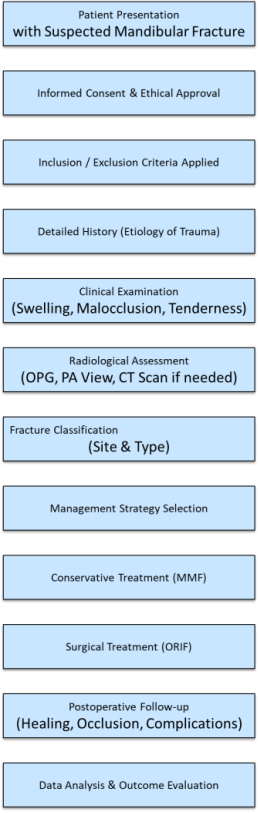
Figure 1. Flow chart of methodology.
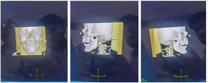
A detailed history was obtained to identify the etiology of trauma, such as road traffic accidents, interpersonal violence, falls, or occupational hazards. Previous studies have highlighted road traffic accidents and interpersonal assaults as the leading causes of mandibular fractures, particularly in developing countries . Clinical examination included inspection and palpation for malocclusion, step deformity, tenderness, swelling, and restricted mandibular movements, as described in earlier classical works.
Radiological assessment was performed using orthopantomogram (OPG) and, when required, posteroanterior mandibular views or computed tomography (CT) for precise localization and classification of fracture lines. Radiological techniques have been shown to be indispensable in accurate diagnosis and treatment planning of mandibular fractures .
Fractures were categorized according to site (symphysis, parasymphysis, body, angle, condyle, or ramus) and type (simple, compound, comminuted, greenstick). Management strategy was selected based on fracture characteristics, displacement, occlusion, and patient factors. Conservative approaches included maxillomandibular fixation (MMF) with arch bars or eyelet wiring, primarily for undisplaced or minimally displaced fractures. Surgical management, involving open reduction and internal fixation (ORIF) with miniplates or reconstruction plates, was employed in displaced, unstable, or multiple fractures, consistent with contemporary protocols described in recent literature .
Postoperative follow-up was undertaken to assess healing, functional recovery, and complications. Data were analyzed to determine the prevalence of etiological factors, fracture patterns, and outcomes of different treatment modalities. The clinicoradiological correlation between findings was emphasized to enhance diagnostic accuracy and guide management .
3. Results and Discussion
The present clinicoradiological study of mandibular fractures in a tertiary care centre provides valuable insights into the demographic distribution, etiology, anatomical sites of involvement, and management strategies adopted for different types of fractures. The findings corroborate with established literature while also reflecting regional trauma trends and evolving treatment protocols.
3.1. Demographic Distribution
The majority of mandibular fractures were reported in males within the age group of 20–40 years. This age distribution is consistent with earlier studies, which have consistently highlighted that young adult males constitute the highest risk group for maxillofacial trauma . Several factors account for this predominance: higher exposure to outdoor activities, increased involvement in high-speed driving, occupational hazards, and social interactions predisposing them to interpersonal violence. In contrast, females and older populations are comparatively less represented, reflecting reduced exposure to such risk factors
Recent studies also confirm this demographic trend. For instance, conducted a global analysis and demonstrated that men in their second and third decades are disproportionately affected by mandibular fractures, with a male-to-female ratio often exceeding 3:1. More recently, Vetter JD et al. (2019) , noted a similar demographic pattern (Figure 2) further underscoring the universality of this trend across both developing and developed countries.
Figure 2. Demographic pattern seen in the patient. Demographic pattern seen in the patient.
3.2. Etiology of Mandibular Fractures
Road traffic accidents (RTAs) emerged as the leading cause of mandibular fractures in this study, followed by accidental falls and interpersonal assaults. The predominance of RTAs is in line with findings reported from various developing nations, where increased motorization, poor road infrastructure, and lack of strict enforcement of traffic regulations significantly contribute to trauma incidence .
In rural and semi-urban settings, motorcycle accidents in particular account for a substantial share of mandibular fractures. Non-compliance with helmet usage and speeding are common contributing factors . Accidental falls were the second most common etiology, often affecting elderly patients and children. Falls in the pediatric group are usually related to play activities, whereas in the elderly, poor balance and systemic conditions predispose to trauma .
Interpersonal violence was identified as another notable cause. This is particularly relevant in younger adult populations, where physical assaults frequently result in mandibular fractures, especially in urban centres. Previous studies from Western countries report a higher proportion of assault-related mandibular fractures compared to RTAs, reflecting differences in social behavior, traffic safety, and cultural patterns . Thus, the current findings align with the epidemiological variations seen worldwide.
3.3. Anatomical Distribution of Fractures
The present study found that the condylar region, parasymphysis, symphysis, and ramus were the most frequently affected anatomical sites. These results are comparable to earlier observations that the mandibular condyle is especially prone to fractures due to its anatomical position and biomechanical properties. Force transmission along the mandible during impact frequently results in condylar involvement, particularly when the chin is struck .
Parasymphysis and symphysis fractures were also commonly observed, which is in agreement with global trends . These regions represent structurally weaker parts of the mandible, especially in the midline, where the bone is relatively thin. Trauma directed to the anterior mandible often results in fractures propagating to the symphysis and parasymphysis.
Ramus fractures, while less common in global reports, were relatively more frequent in this study. This variation may be attributed to specific trauma patterns, particularly high-energy RTAs, where lateral impacts can transfer significant force to the ramus. Moreover, the increased reporting of ramus fractures may also reflect improved diagnostic accuracy with advanced imaging modalities such as CT scans, which were not routinely available in older studies.
3.4. Management of Mandibular Fractures
The treatment strategies adopted in this study highlight the clinical judgment required in balancing functional outcomes, patient comfort, and resource availability. Displaced fractures were predominantly treated with open reduction and internal fixation (ORIF) using titanium miniplates. This approach ensured anatomical realignment, early restoration of function, and reduced morbidity associated with prolonged maxillomandibular fixation (MMF). The introduction of Champy’s principles of miniplate osteosynthesis has revolutionized mandibular fracture management by providing stable fixation while preserving periosteal blood supply .
Numerous studies support ORIF as the gold standard for displaced mandibular fractures. For example, a systematic review by highlighted the superior outcomes of miniplate fixation in terms of occlusal stability, masticatory efficiency, and reduced healing time. More recent literature also emphasizes the advantages of rigid fixation, including early mobilization, shorter hospital stays, and lower incidence of complications such as malunion and temporomandibular joint dysfunction .
On the other hand, conservative management was successfully employed in non-displaced fractures and pediatric cases. Intermaxillary fixation (IMF) with arch bars, eyelet wiring, or elastics, along with dietary modifications, proved sufficient to achieve satisfactory healing in these patients. The rationale for conservative management in children lies in their greater osteogenic potential and the risks associated with rigid fixation in developing bones, such as growth disturbances and tooth bud damage.
This approach aligns with earlier findings by , who advocated conservative treatment in pediatric fractures Table 1). Recent reviews continue to emphasize the importance of minimizing surgical intervention in children to preserve mandibular growth potential .
3.4.1. Inclusion & Exclusion Criteria
1) Inclusion: All age groups, both genders, radiologically confirmed fractures.
2) Exclusion: Pathological fractures, previous mandibular surgery, inadequate records.
3.4.2. Demographics & Etiology
1) Majority: males 20–40 yrs
2) Leading cause: Road traffic accidents (58%)
3) Others: Accidental falls (25%), interpersonal assaults (17%)
Table 1. Conservative treatment in pediatric fractures. Conservative treatment in pediatric fractures. Conservative treatment in pediatric fractures.

Category

Subcategory

Percentage (%)

Age Group (years)

20–40

62%

0–19

18%

41 & above

20%

Etiology

Road Traffic Accidents

58%

Accidental Falls

25%

Interpersonal Assaults

17%

Fracture Site

Condylar Region

34%

Parasymphysis

28%

Symphysis

22%

Ramus

16%

Treatment Modality

ORIF (Miniplates)

61%

Conservative (IMF/Dietary)

39%

3.5. Outcome Evaluation
The outcomes observed in this study were generally favorable, with patients treated by ORIF achieving early restoration of mandibular function and occlusion. Conservative treatment also demonstrated satisfactory healing, particularly in pediatric and minimally displaced fractures . Few complications, such as infection, malocclusion, or delayed union, were encountered, reflecting both appropriate case selection and adherence to treatment protocols .
The combination of clinical and radiological evaluation was pivotal in guiding management and ensuring accurate diagnosis. The correlation between clinical findings such as step deformity, malocclusion, and restricted mandibular movements with radiological evidence enhanced diagnostic confidence and aided in surgical planning.
3.6. Comparison with Literature
The findings of this study broadly correspond to earlier reports while also highlighting certain regional differences. The predominance of RTAs mirrors observations from other developing nations . However, the relatively higher incidence of ramus fractures underscores the importance of local trauma mechanisms and warrants further investigation.
Management strategies adopted here reflect global trends, with ORIF increasingly being considered the standard of care for displaced fractures, while conservative approaches remain relevant for select cases. This balanced approach ensures optimal functional and esthetic outcomes while minimizing complications.
4. Conclusions
This clinicoradiological study reaffirms the demographic, etiological, and anatomical patterns of mandibular fractures while underscoring the importance of tailored management strategies. Young adult males remain the most affected group, with RTAs as the predominant cause. Condylar, parasymphysis, and symphysis fractures are the most frequent, and their management requires judicious selection between ORIF and conservative methods.
The study emphasizes the indispensable role of clinicoradiological correlation in diagnosis and treatment planning. Furthermore, the favorable outcomes observed underscore the effectiveness of adopting evidence-based treatment protocols in a tertiary care setting.
Abbreviations

RTA

Road Traffic Accident

OPG

Orthopantomogram

CT

Computed Tomography

ORIF

Open Reduction and Internal Fixation

MMF

Maxillomandibular Fixation

IMF

Intermaxillary Fixation

ENT

Ear, Nose and Throat

TMJ

Temporomandibular Joint

Conflicts of Interest
The authors declare no conflicts of interest.
References
[1] Rai A, Karwal V, Nigam S, Saxena A, Sharma M. Outcome study of mandibular fractures treated by surgical stabilization with plates and screws. Cureus. 2024; 16(4): e58561.
[2] Oxford M, Tucker J, Goldenberg D, King TS, Ziai K, Henry C, Lighthall JG. Comparison of pediatric and adult mandibular fractures: identifying differences in presentation and management using a national trauma database. J Craniofac Surg. 2025; 36(2): 461–464.
[3] Roccia F, Cena P, Cremona G, Garzino Demo P, Sobrero F. Characteristics and surgical management of bilateral body mandibular fractures: a 23-year experience. J Clin Med. 2025; 14(1): 160.
[4] Mudawi AIAA. Mandibular fractures: a five-year retrospective review. Egypt J Health Care. 2025; 16(3): 845–852.
[5] Kawilarang B, Sanjaya IGPH, Hariantana AR. A five-year epidemiological analysis of mandible fractures in Bali. Jurnal Plastik Rekonstruksi. 2025; 12(1): 7–13.
[6] Khan W, Syed K, Khan S, Safwan AA, Saboor A, Khan A, Rahim M. Outcome of open reduction and internal fixation using one titanium plate and arch bar for mandibular fractures in symphyseal and parasymphyseal region. J Gandhara Med Dent Sci. 2025; 12(4): 53–57.
[7] Vetter JD, Topazian RG, Goldberg MH, Smith DG. Facial fractures occurring in a medium-sized metropolitan area: recent trends. Int J Oral Maxillofac Surg. 2019; 48(11): 1411–1418,
[8] Boffano P, Kommers SC, Roccia F, Forouzanfar T. Mandibular trauma: a two-century review. Int J Oral Maxillofac Surg. 2015; 44(12): 1389–1399.
[9] Kumar N, Yadav SK, Tandon R, Chandra A. Management of pediatric mandibular fractures: a review. Natl J Maxillofac Surg. 2020; 11(1): 3–9.
[10] Champy M, Loddé JP, Schmitt R, Jaeger JH, Muster D. Mandibular osteosynthesis by miniature screwed plates via a buccal approach. J Maxillofac Surg. 1976; 4(1): 14–21.
[11] Al-Ahmed HE, Jaber MA, Fanas SH, Karas M. The pattern of maxillofacial fractures in Sharjah, United Arab Emirates: a review of 230 cases. Oral Surg Oral Med Oral Pathol Oral Radiol Endod. 2004; 98(2): 166-170.
[12] Lee KH. Global trends in maxillofacial fractures. Craniomaxillofac Trauma Reconstr. 2008; 1(1): 31-36.
[13] Rowe NL, Killey HC. Fractures of the Facial Skeleton. Edinburgh: Churchill Livingstone; 1968.
[14] Ellis E, Moos KF, el-Attar A. Ten years of mandibular fractures: an analysis of 2,137 cases. Oral Surg Oral Med Oral Pathol. 1985; 59(2): 120-129.
[15] F. Roccia, P. Cena, G. Cremona, P. Garzino Demo, and F. Sobrero, “Characteristics and Surgical Management of Bilateral Body Mandibular Fractures: A 23?Year Experience,” J. Clin. Med., vol. 14, no. 1, article 160 (pp. 1–15), 2025.
[16] Chrcanovic BR. Factors influencing the incidence of maxillofacial fractures, Oral Maxillofac Surg. 2012; 16(1): 3–17.
[17] Haug RH, Prather J, Indresano AT. An epidemiologic survey of facial fractures, J Oral Maxillofac Surg. 1990; 48(9): 926–932.
[18] Madison Oxford, Jacqueline Tucker, Dana Goldenberg, Tonya S. King, Kasra Ziai, Cathy Henry, and Jessyka G. Lighthall, “Comparison of Pediatric and Adult Mandibular Fractures: Identifying Differences in Presentation and Management Using a National Trauma Database,” J. Craniofac. Surg., vol. 36, no. 2, pp. 461–464, 2025.
[19] Brasileiro BF, Passeri LA. Epidemiological analysis of maxillofacial fractures in Brazil, J Oral Maxillofac Surg. 2006; 64(7): 1019–1024.
[20] Dongas P, Hall GM. Mandibular fracture patterns in Tasmania, Australia, Aust Dent J. 2002; 47(2): 131–137.
[21] Kroon FH, Mathisson M, Cordey JR, Rahn BA. The use of miniplates in mandibular fractures, J Craniomaxillofac Surg. 1991; 19(5): 199–204.
[22] Adeyemo WL, Ladeinde AL, Ogunlewe MO, James O. Trends and characteristics of oral and maxillofacial injuries in Nigeria: a review of the literature. Head Face Med. 2011; 7: 6.
[23] Posnick J. C. Pediatric facial fractures: evolving patterns of treatment, J Oral Maxillofac Surg. 2000; 58(2): 136–147.
[24] Ellis E, Walker L. Treatment of mandibular angle fractures using one noncompression miniplate, J Oral Maxillofac Surg. 1996; 54(7): 864–871.
[25] Ellis E. Treatment methods for fractures of the mandibular angle, Int J Oral Maxillofac Surg. 1999; 28(4): 243–252. HYPERLINK "
Cite This Article
  • APA Style

    Gujrathi, A. B., Paikrao, Y. M., Rewatkar, A. K. (2026). Clinicoradiological Study of Mandibular Fractures in Tertiary Care Centre. International Journal of Clinical Oral and Maxillofacial Surgery, 12(1), 1-6. https://doi.org/10.11648/j.ijcoms.20261201.11

    Copy | Download

    ACS Style

    Gujrathi, A. B.; Paikrao, Y. M.; Rewatkar, A. K. Clinicoradiological Study of Mandibular Fractures in Tertiary Care Centre. Int. J. Clin. Oral Maxillofac. Surg. 2026, 12(1), 1-6. doi: 10.11648/j.ijcoms.20261201.11

    Copy | Download

    AMA Style

    Gujrathi AB, Paikrao YM, Rewatkar AK. Clinicoradiological Study of Mandibular Fractures in Tertiary Care Centre. Int J Clin Oral Maxillofac Surg. 2026;12(1):1-6. doi: 10.11648/j.ijcoms.20261201.11

    Copy | Download

  • @article{10.11648/j.ijcoms.20261201.11,
      author = {Atishkumar Balajirao Gujrathi and Yogesh Madhaorao Paikrao and Awantika Kishor Rewatkar},
      title = {Clinicoradiological Study of Mandibular Fractures in Tertiary Care Centre},
      journal = {International Journal of Clinical Oral and Maxillofacial Surgery},
      volume = {12},
      number = {1},
      pages = {1-6},
      doi = {10.11648/j.ijcoms.20261201.11},
      url = {https://doi.org/10.11648/j.ijcoms.20261201.11},
      eprint = {https://article.sciencepublishinggroup.com/pdf/10.11648.j.ijcoms.20261201.11},
      abstract = {The mandible, being one of the most prominent and mobile bones of the facial skeleton, is highly susceptible to traumatic injuries. Mandibular fractures represent a significant proportion of maxillofacial trauma cases, often resulting from road traffic accidents, assaults, falls, or occupational injuries. The present study, titled “Clinicoradiological Study of Mandibular Fractures in Tertiary Care Centre”, aims to analyze in detail the etiology, clinical presentation, radiological patterns, and types of mandibular fractures encountered in a tertiary care setting in the span of two years (October 2023 - October 2025). Special emphasis is placed on the diagnostic role of clinicoradiological correlation in accurate localization and classification of fractures. Furthermore, the study evaluates different management modalities, ranging from conservative approaches such as closed reduction and maxillomandibular fixation to surgical interventions like open reduction and internal fixation. By comparing outcomes of these treatment strategies, the study seeks to highlight the practical considerations, challenges, and effectiveness of both conservative and surgical methods in routine clinical practice.},
     year = {2026}
    }
    

    Copy | Download

  • TY  - JOUR
    T1  - Clinicoradiological Study of Mandibular Fractures in Tertiary Care Centre
    AU  - Atishkumar Balajirao Gujrathi
    AU  - Yogesh Madhaorao Paikrao
    AU  - Awantika Kishor Rewatkar
    Y1  - 2026/01/29
    PY  - 2026
    N1  - https://doi.org/10.11648/j.ijcoms.20261201.11
    DO  - 10.11648/j.ijcoms.20261201.11
    T2  - International Journal of Clinical Oral and Maxillofacial Surgery
    JF  - International Journal of Clinical Oral and Maxillofacial Surgery
    JO  - International Journal of Clinical Oral and Maxillofacial Surgery
    SP  - 1
    EP  - 6
    PB  - Science Publishing Group
    SN  - 2472-1344
    UR  - https://doi.org/10.11648/j.ijcoms.20261201.11
    AB  - The mandible, being one of the most prominent and mobile bones of the facial skeleton, is highly susceptible to traumatic injuries. Mandibular fractures represent a significant proportion of maxillofacial trauma cases, often resulting from road traffic accidents, assaults, falls, or occupational injuries. The present study, titled “Clinicoradiological Study of Mandibular Fractures in Tertiary Care Centre”, aims to analyze in detail the etiology, clinical presentation, radiological patterns, and types of mandibular fractures encountered in a tertiary care setting in the span of two years (October 2023 - October 2025). Special emphasis is placed on the diagnostic role of clinicoradiological correlation in accurate localization and classification of fractures. Furthermore, the study evaluates different management modalities, ranging from conservative approaches such as closed reduction and maxillomandibular fixation to surgical interventions like open reduction and internal fixation. By comparing outcomes of these treatment strategies, the study seeks to highlight the practical considerations, challenges, and effectiveness of both conservative and surgical methods in routine clinical practice.
    VL  - 12
    IS  - 1
    ER  - 

    Copy | Download

Author Information
  • Abstract
  • Keywords
  • Document Sections

    1. 1. Introduction
    2. 2. Methodology
    3. 3. Results and Discussion
    4. 4. Conclusions
    Show Full Outline
  • Abbreviations
  • Conflicts of Interest
  • References
  • Cite This Article
  • Author Information
  • Table 1

    Table 1. Conservative treatment in pediatric fractures. Conservative treatment in pediatric fractures.